Abdominal Anatomy - Axial Muscles Of The Abdominal Wall And Thorax Anatomy Physiology / The abdominal body wall and the pelvis are the topics of this week.
Abdominal Anatomy - Axial Muscles Of The Abdominal Wall And Thorax Anatomy Physiology / The abdominal body wall and the pelvis are the topics of this week.. Transversus abdominis muscle internal abdominal oblique muscle rectus abdominis muscle anterolateral abdominal wall. Sciency root words make anatomical parts harder to memorize. Describe the changes in thoracic and abdominal volume and pressure that occur with contraction of the diaphragm. Abdominal wall anatomy that is clinically pertinent to the surgeon, focusing primarily on the structures of the anterior abdominal wall, will be reviewed. Windham was previously a surgical.
We'll identify as many organs as we can. Simple, easy notes for quick revision of important questions. Divided into 9 regions by two vertical and two horizontal imaginary planes. Athletic injuries of the lateral abdominal wall: The abdomen (colloquially called the belly, tummy, midriff or stomach) is the part of the body between the thorax (chest) and pelvis, in humans and in other vertebrates.
The abdomen contains all of the digestive.
Abdominal wall anatomy that is clinically pertinent to the surgeon, focusing primarily on the structures of the anterior abdominal wall, will be reviewed. Simple, easy notes for quick revision of important questions. We're going to take apart a plastic anatomy model and see what we can find in the abdomen. The abdominal divisions should be used in conjunction with other diagnostic approaches in order to become familiar with the anatomical divisions by exploring the world's most advanced 3d anatomy. Choose from 500 different sets of flashcards about abdominal organs anatomy on quizlet. Sectional anatomy the sonographer must have figure 5: The abdomen (colloquially called the belly, tummy, midriff or stomach) is the part of the body between the thorax (chest) and pelvis, in humans and in other vertebrates. The anterior abdominal wall (figs. Introduction to sonographic abdominal anatomy. Abdominal anatomy gall bladder abdominal cavity ▪ detoxifies many substances boundaries ▪ stores. Transverse mesocolon attached the duodenum to the posterior abdominal wall. • in this module, we will explore basic abdominal anatomy identifiable with common imaging modalities. Abdominal surface anatomy can be described when viewed from in front of the abdomen in 2 ways:
Simple, easy notes for quick revision of important questions. But with the use of smart technology, you can learn faster and master abdomen anatomy in no time! Abdominal surface anatomy can be described when viewed from in front of the abdomen in 2 ways: Abdominal wall anatomy that is clinically pertinent to the surgeon, focusing primarily on the structures of the anterior abdominal wall, will be reviewed. Windham was previously a surgical.

Divided into 9 regions by two vertical and two horizontal imaginary planes.
• in this module, we will explore basic abdominal anatomy identifiable with common imaging modalities. Laterally by the midaxillary line. Unit three — abdominal organs, pelvis & lower limb. Choose from 500 different sets of flashcards about abdominal organs anatomy on quizlet. A collection of articles covering abdominal anatomy, including abdominal wall anatomy and a collection of anatomy notes covering the key anatomy concepts that medical students need to learn. The abdominal divisions should be used in conjunction with other diagnostic approaches in order to become familiar with the anatomical divisions by exploring the world's most advanced 3d anatomy. Divided into 9 regions by two vertical and two horizontal imaginary planes. A good amount of area is covered by the abdominal wall. Introduction to sonographic abdominal anatomy. We'll identify as many organs as we can. We created an anatomical atlas of abdominal and pelvic ct which is an interactive tool for studying the conventional anatomy of the normal structures based on a multidetector computed tomography. Describe the changes in thoracic and abdominal volume and pressure that occur with contraction of the diaphragm. Transverse mesocolon attached the duodenum to the posterior abdominal wall.
Review of anatomy and mr imaging appearance. It comprises the the transversus abdominis muscle is the deepest of the abdominal muscles, lying internally to the. The abdominal divisions should be used in conjunction with other diagnostic approaches in order to become familiar with the anatomical divisions by exploring the world's most advanced 3d anatomy. Introduction to sonographic abdominal anatomy. Abdomen anatomy mcqs a total of 138 mcqs that cover the anatomy of abdomen region 7.
Laterally by the midaxillary line.
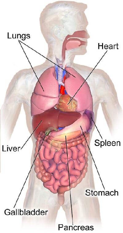
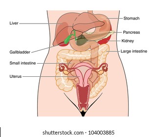
The abdomen contains all of the digestive. Review of anatomy and mr imaging appearance. The abdomen contains many vital organs: Abdominal surface anatomy can be described when viewed from in front of the abdomen in 2 ways: A collection of articles covering abdominal anatomy, including abdominal wall anatomy and a collection of anatomy notes covering the key anatomy concepts that medical students need to learn. Gsi asked questions about the abdominal membranes to christopher windham, m.d. Transverse mesocolon attached the duodenum to the posterior abdominal wall. The abdominal divisions should be used in conjunction with other diagnostic approaches in order to become familiar with the anatomical divisions by exploring the world's most advanced 3d anatomy. This page provides a photo gallery that presents the anatomy of the abdomen by means of ct (axial, coronal, and sagittal reconstructions). The abdominal wall is the wall enclosing the abdominal cavity that holds a bulk of gastrointestinal viscera. • in this module, we will explore basic abdominal anatomy identifiable with common imaging modalities. Abdomen anatomy mcqs a total of 138 mcqs that cover the anatomy of abdomen region 7. Athletic injuries of the lateral abdominal wall:


Komentar
Posting Komentar